
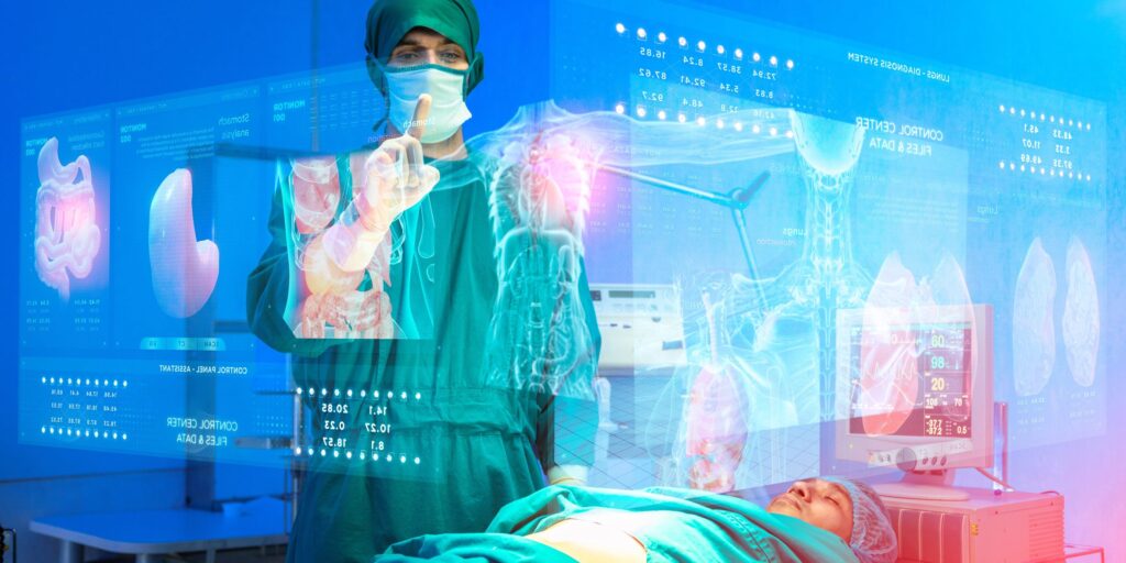
Augmented Reality, with its ability to overlay digital content onto the physical world, has opened new frontiers in how we interact with data, systems, and one another. In the healthcare sector, this forward-thinking technology is increasingly considered a potential game-changer. By allowing medical professionals to bring complex data into a three-dimensional space, it offers opportunities to transform patient examinations, surgical procedures, and medical education. Beyond the initial excitement, there is a growing realisation among clinicians and researchers that Augmented Reality can significantly enhance their ability to see, interpret, and understand the intricate patterns found in medical imaging data.
Viewing diagnostic imagery such as MRI scans, X-rays, and CT scans in a two-dimensional format has long been the standard. Although these methods have made rapid strides in clarity and resolution, translating flat images into three-dimensional anatomical and pathological contexts remains a challenge for many healthcare professionals. Augmented Reality offers a means to bridge this gap. It can layer three-dimensional models of organs, tissues, and even microscopic structures within the doctor’s or surgeon’s immediate surroundings. Yet the practicalities of converting raw medical imaging data into such interactive experiences are far from trivial. This is where advanced data processing, high-performance visualisation technologies, and real-time rendering engines come into play, enabling the projection of detailed anatomical models without sacrificing speed or clarity.
Choosing the right visualisation tools like JavaScript charts
One developer from SciChart advises that choosing the right visualisation tools like JavaScript charts and optimising data throughput is crucial for building high-performance AR solutions in healthcare. They emphasise that rendering speed and clarity have to strike a delicate balance. It is not enough to have a robust engine if the display lags behind the user’s physical movements. Equally, data density can impact how effective visual overlays can be, so working with optimised libraries—whether that involves JavaScript charts or native modules for medical imaging—can significantly enhance the usability of AR applications in a clinical setting.
The Growth of Augmented Reality in Healthcare
Augmented Reality has seen a particularly visible adoption curve over the last decade, propelled by an increasing desire to interact with digital content more intuitively. Major technology companies have introduced AR-capable devices and software frameworks that promise to revolutionise how we perceive the world. In healthcare, the interest in AR is not motivated by novelty alone but rather by its potential to address critical pain points in patient care and medical research. Surgeons, for instance, have started to experiment with headsets that display layered 3D models of patient anatomy during surgical procedures. This overlay can help with targeting tumours, navigating blood vessels, or placing implants in a more precise manner.
While some of these AR applications remain in experimental phases, others are beginning to gain traction. Hospitals in certain regions have adopted AR-based training modules that allow medical students and trainees to visualise a complete anatomical structure over a life-sized mannequin or even a volunteer. The promise is evident: AR creates interactive learning experiences that are immersive and can drastically shorten the time needed to understand complex anatomical relationships. However, the surge in usage also highlights the importance of high-fidelity software frameworks and rigorous data visualisation standards. Not only must these tools handle large data sets with minimal latency, they also need to integrate seamlessly with existing medical imaging formats and protocols.
Understanding Complex Medical Imaging Data
Medical imaging comes in many forms, each possessing its strengths and limitations. MRI scans provide detailed images of soft tissue; X-rays are suitable for visualising bone and certain other dense structures; CT scans offer cross-sectional views that can be reconstructed into three-dimensional images. Within each of these modalities, there is a wealth of detail that demands efficient data processing to be represented accurately. From raw pixel data to segmented 3D volumes, the transformation pipeline is highly specialised and requires a thorough understanding of image acquisition techniques.
In traditional two-dimensional viewing, radiologists examine slice-by-slice images on a computer screen. Although decades of practice have enabled radiologists to visualise three-dimensional forms mentally, the cognitive burden is not insignificant. Augmented Reality helps alleviate some of this strain by generating three-dimensional reconstructions and enabling the observer to walk around a model, rotate it in mid-air, or adjust its transparency to reveal hidden layers of tissue. The result is a more intuitive grasp of spatial relationships and the potential for improved diagnostic accuracy. Yet this transformation of raw data into a 3D structure is technically complex. Visual artefacts, rendering performance, and image segmentation inaccuracies can impair the reliability of AR overlays, thereby requiring advanced algorithms, robust hardware, and stringent validation protocols.
Challenges in Visualising Medical Imaging Data
Ensuring that AR experiences are medically reliable is a challenge involving multiple factors. First, the data itself must be of high resolution. Medical imaging devices such as MRI and CT scanners can generate an immense volume of slices and views. The size of these data sets can be quite large, straining even high-end computers when attempting to create real-time interactive graphics. Second, the image processing pipeline must convert this raw data into meaningful 3D shapes. This can involve segmenting out organs or lesions from surrounding tissues, then accurately rendering them within the AR system. Misalignment or misregistration in segmentation could lead to incorrect overlays, diminishing the trust clinicians place in these tools.
In addition, the computing resources required for Augmented Reality go beyond just converting images into polygons or point clouds. The system must track the user’s viewpoint in real time, update the rendered image accordingly, and display it at a comfortable frame rate for the user. This necessitates low-latency operations, sophisticated head tracking, and efficient memory management. Developers often rely on advanced rendering engines and software libraries that can take advantage of parallel processing on graphics cards. Furthermore, the AR display device itself must be accurate. Differences in lens refractions or user-specific eyesight corrections can introduce discrepancies between the real and virtual overlays. Sub-millimetre accuracy in medical procedures can be the difference between success and complication, so AR solutions need to be built with these stringent standards in mind.
Augmented Reality as a Solution
Despite the inherent complexities, Augmented Reality stands out as a powerful method of delivering complex medical imaging data to the point of care. Traditional displays are constrained by their two-dimensional nature, and while three-dimensional reconstructions on a monitor can approximate depth using shading and perspective, AR allows a more holistic view. Clinicians can not only look at organ structures from multiple angles but also measure distances or angles by literally moving around the virtual model. This capability is particularly potent in pre-surgical planning. Surgeons can study a patient-specific model of an organ, identify potential complications, and plan incisions or instrument pathways well in advance.
Beyond preoperative planning, there is the potential for real-time intraoperative guidance. In some systems, the patient’s real-time imaging data can be projected over their actual body, enabling surgeons to pinpoint the exact location of an underlying tumour. The healthcare team can then cross-reference the data with actual patient anatomy without shifting their gaze to a separate display. This form of synchronous visualisation has been linked to shortened operating times and potentially more accurate outcomes, although rigorous clinical trials are still underway to substantiate these claims.
Importance of Real-Time Data Visualisation Tools
Underpinning much of the progress in AR-based medical imaging is the capacity to visualise data in real time. The healthcare environment can be fast-paced, and professionals often need immediate access to new information. In the context of surgeries or emergency care, even a few seconds of delay in updating the visual overlay might render the tool unusable. Real-time visualisation tools, therefore, are at the forefront of AR adoption. These tools typically rely on specialised software libraries that can manage and render massive data sets with minimal latency. Implementing advanced features such as occlusion handling, edge detection, or lighting corrections requires high levels of compute power, particularly if large 3D volumes are being displayed.
Some developers turn to frameworks that were initially designed for other industries, adapting them to medical settings. For example, a high-performance JavaScript charting library can be used in web-based interfaces that clinicians access on tablets or AR headsets. While such libraries are more commonly associated with financial or scientific data, the underlying rendering engines can be repurposed to handle advanced medical visualisation tasks. In certain cases, the ability to integrate real-time updates—such as changes in physiological parameters—into an AR view can be facilitated by data streaming via secure servers. This might involve the use of JavaScript charts to provide auxiliary information, like real-time heart rate or blood pressure trends, layered alongside the 3D anatomical model. If done well, this composite of data feeds can significantly enhance clinical decision-making.
Potential Applications of AR in Medical Imaging
The scope of Augmented Reality in medical imaging spans both diagnostics and intervention. During diagnostic consultations, radiologists can display a three-dimensional representation of an MRI scan right on their desk, guiding patients through complex findings in an engaging manner. Patients who can see a virtual replica of their anatomy might have a clearer understanding of their condition, leading to higher adherence to treatment plans. AR can also be used to compare current imaging results with previous scans side by side in 3D, aiding in the assessment of disease progression or the effectiveness of treatments.
In operating theatres, AR can offer assistance in surgical navigation, particularly when the precise location of critical structures is paramount. By aligning the patient’s imaging data with their physical body, surgeons may reduce reliance on guesswork and avoid vital nerves or blood vessels. Another area where AR could flourish is in remote consultations and telemedicine. Experts from one location can see the same 3D model overlay that the local physician is viewing, offering real-time advice without the need to physically travel. This blending of advanced visualisation and communication technologies has wide-ranging implications for global healthcare, particularly in remote or under-resourced areas.
Educational environments also stand to benefit. Medical students often rely on cadavers and textbooks to understand how organs fit together within the human body. AR adds a digital overlay to actual models, letting students peel back layers, isolate specific organs, and view pathological variations in a detailed manner. This level of interaction could improve both retention and comprehension. Similarly, practising physicians can use AR training modules to rehearse rare procedures before attempting them on actual patients. These sorts of simulated environments provide an extra layer of safety and preparation.
Security and Ethical Considerations
AR solutions in healthcare, like all digital systems, bring with them the responsibility to ensure patient privacy and data security. Medical imaging data often contains sensitive personal information, and any platform that handles this data must comply with relevant regulations and standards. Encryption of data streams becomes critical when loading high-resolution medical scans into an AR application, especially if the data is transmitted across networks. Additionally, the storage of processed 3D models must be done in compliance with patient confidentiality protocols.
Another layer of responsibility involves verifying the accuracy of any AR-guided intervention. If a system misrepresents the location of a lesion, the consequences could be dire. Therefore, thorough testing, calibration, and certification processes are essential. Clinicians and software developers must work together to validate each step of the pipeline, from the acquisition of raw images to the rendering of 3D anatomy in a headset. Ethical debates also arise around the potential for AR to shift liability. If a surgeon relies heavily on an AR overlay that turns out to be flawed, how is responsibility assigned? Addressing such questions will require robust policy frameworks and collaborative efforts from healthcare authorities, technology providers, and legal experts.
The Role of JavaScript Charts in Data Integration
While the visualisation of complex imaging data in AR environments often relies on sophisticated 3D rendering engines, other forms of clinical data must also be integrated to provide a comprehensive overview. This is where JavaScript charts can become a supporting component in the overall architecture of an AR medical solution. By nature, charts present trends and relationships in a more traditional two-dimensional context. Clinicians may wish to view blood test results, heart rate monitoring over time, or changes in tumour size across multiple scans. JavaScript charts can quickly be embedded in the user interface, offering an easily interpretable snapshot of these data sets. Their flexibility and performance capabilities in web-based environments make them a logical choice for scenarios where multiple data streams converge into a single application.
Even though the key spotlight in augmented medical imaging is often on volumetric rendering and complex overlays, supplementary data visualisation is no less critical. The ability to observe real-time metrics while inspecting a 3D anatomical structure can assist clinicians in making immediate, well-informed decisions. That is why so many developers are adopting frameworks that not only generate advanced 3D models but also integrate real-time charting libraries that handle line charts, bar charts, or scatter plots seamlessly. When orchestrated effectively, the layering of interactive 3D models with well-designed charts can heighten the depth of insight clinicians glean from patient data.
Commentary on Emerging Technologies
An intriguing development arises from the growth of cloud computing and the adoption of frameworks such as React. Some teams combine React charts with augmented visual overlays to offer dynamic user interfaces. Although AR primarily deals with 3D geometry, there is room for more conventional data visualisation to appear as floating panels or supplementary displays within the user’s field of view. By harnessing the strengths of these libraries, clinical teams can potentially experience a comprehensive interface where volumetric imaging data, laboratory results, and patient history co-exist in one fluid environment. While still a work in progress, the convergence of AR and interactive charting libraries promises to refine clinical workflows and augment the value provided by next-generation healthcare applications.
Future Outlook
The future of Augmented Reality in healthcare holds great promise. As hardware becomes more compact and computational power grows, AR devices will be capable of rendering highly detailed 3D structures with even lower latency. Advances in machine learning and artificial intelligence will streamline the segmentation and interpretation of medical images, making the conversion from a flat scan to an interactive 3D model more accurate and faster. The continued integration of real-time data visualisation tools, including JavaScript charts, within AR environments will further enhance the holistic view clinicians can have on patient data. Automated anomaly detection, personalised anatomical modelling, and predictive analytics are all within reach, possibly giving clinicians the ability to foresee complications before they manifest.
The technology might also move beyond handheld or head-mounted devices to room-scale or even holographic projections. In these scenarios, multiple practitioners could gather around a shared 3D representation of patient data, interacting with it from different angles and collaborating in a more immersive way. The links between telemedicine and AR are also likely to strengthen. Remote experts could participate in live surgeries, manipulating AR overlays in real time to guide less experienced clinicians. This could dramatically improve the standard of care in locations that lack specialised expertise, bridging geographical disparities that have historically been difficult to address. While these visions of the future are inspiring, they hinge on robust technological ecosystems and the willingness of healthcare stakeholders to adapt longstanding practices.
Conclusion
Augmented Reality represents a leap forward in the way complex medical imaging data can be visualised and employed in clinical care. By shifting from flat, two-dimensional displays to immersive, three-dimensional reconstructions, it opens a multitude of possibilities in diagnostics, surgical navigation, and medical education. The path to widespread adoption is not without obstacles, encompassing substantial technical demands, ethical considerations, and the need for stringent verification. Nonetheless, as hardware capabilities evolve and software frameworks mature, more healthcare institutions are likely to find AR-based solutions both feasible and beneficial.
The role of supplemental technologies, such as JavaScript charts, is equally significant. These tools contribute to the unified visual environment by providing straightforward ways to present real-time measurements and historical data. Pairing high fidelity 3D imaging with charts that display trends and numeric values can help clinicians arrive at more informed decisions. As research continues and more clinical trials validate the efficacy of AR-guided interventions, the medical community will likely integrate these solutions further into everyday practice. For now, the industry stands on the cusp of an exciting evolution, one in which augmented visualisations guide hands in the operating theatre and bring a new level of clarity to patient consultations, ultimately improving care outcomes and enhancing the efficiency of modern healthcare systems.
READ MORE: Prostavive Colibrim: A Natural Solution for Prostate Health and Vitality